
Animal models have historically been the primary means through which the human body can be modelled. Mice, pigs, and other animals have all provided useful insights into how we would respond to therapeutics and where diseases originate.
Yet more and more therapeutic candidates are failing to get past clinical trials despite strong results from animal models. Combined with a desire to ensure animal welfare, regulatory boards worldwide encouraged scientists to consider other methods to model human anatomy and pathophysiology. Known as New Approach Methodologies (NAMs), they comprise new testing methods — organoids, computational tools, and other complex systems — that mimic human biology without the need for animal models.
Among these models are zebrafish. What’s interesting about them is that they’ve already been used for decades because they can develop quickly, have transparent embryos for development biology research, and are highly similar to humans genetically. While it takes additional funds to run a zebrafish colony, the ability to screen these embryos for morphological features translates well to screening other kinds of cells.
It’s that feature that Frank Bonnet, CEO and co-founder of Bionomous, capitalized on when developing the Sortivo. His inventive approach to 3D tissue sorting, borne from his research into zebrafish, helped Bionomous win the New Product Award at SLAS Europe 2026. I had a chance to sit down with him to talk about his approach to screening 3D models and how it can accelerate preclinical research.
Learn more through the interview below.
PN: You initially studied fish behaviour by building robot fish. What were you studying, and how did it inspire you to start Bionomous?
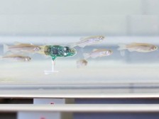
FB: It was during my PhD years that I constructed a robot fish the size of a zebrafish (Figure 1). I built it to study schooling behavior in zebrafish as part of a larger European research project investigating animal collective behavior through bio-inspired robotic systems.
As I developed the robotic fish, I worked directly with biologists who used zebrafish as a mode for biological research. It was during those collaborations that I discovered how powerful zebrafish are not only for studying animal behavior, but also as a model for understanding human diseases and accelerating drug discovery. At the same time, I saw many of the practical challenges researchers faced, particularly the labour-intensive and repetitive manual handling of zebrafish embryos and larvae.
I have always striven to develop technologies that improve researchers’ daily work. Recognizing this unmet need, I began designing an automated solution for handling these biological samples. That work ultimately became the foundation of the technology behind Bionomous.
PN: Zebrafish are among a growing group known as new approach methodologies (NAMs). As regulatory boards and research organizations seek to move away from animal testing, why should scientists consider integrating these approaches earlier in the discovery pipeline?
FB: Traditional animal models, particularly mice, are both ethically and financially costly. It takes substantial time and resources to establish an animal testing facility. Large numbers of animals are also sacrificed just to obtain meaningful preclinical data.
Despite that effort, over 90% of drug candidates that show promising results in preclinical animal studies ultimately fail during human clinical trials. This has compromised the predictive value of these models for human biology and highlighted the need for complementary models that better reflect human physiology. That doesn’t mean that animal models have lost their use in preclinical research. It just means that we need to integrate other models earlier in the drug discovery pipeline.
NAMs provide that bridge for earlier preclinical research. Human cell-based models, including organoids and other advanced in vitro systems, can often be derived from patient cells or engineered to closely reproduce specific tissues or disease states. As a result, they can provide more relevant biological information earlier in the drug discovery process, while reducing costs, shortening development timelines, and minimizing animal use.
PN: Zebrafish is an example of a NAM, but with other NAMs such as organoids available, has that affected how likely researchers are to use zebrafish for drug development?
FB: Many of our customers who have traditionally worked with zebrafish are now considering or have also already incorporated organoids into their research. At the same time, those researchers are using organoids as a complementary tool rather than replacing zebrafish outright. On the one hand, organoids provide highly relevant human-specific models that can answer different biological questions. On the other hand, zebrafish are complete living organisms, so researchers can study complex physiological processes such as development, organ interactions, and whole-body responses.
PN: Since starting Bionomous, you’ve launched the Sortivo, a machine that screens, sorts, and plates organoids and fish larvae. One feature that particularly sticks out is its capability to conduct 360o imaging in brightfield and fluorescence formats. What happens to the sample during the imaging step, and how does observing the sample from multiple angles expand what scientists can evaluate beyond existing fluorescence-based sorting methods?
FB: The sample is transferred into an imaging chamber where it can be oriented and imaged from multiple perspectives using either brightfield or fluorescence imaging. This enables the reconstruction of a 3D model of the sample.
For zebrafish larvae, this allows researchers to visualize organs from different viewpoints. For organoids, it enables the observation of cellular structures and growth patterns throughout the entire volume, including the spatial distribution of specific cell populations.
Performing this type of multi-perspective imaging manually would typically require hours of work in a dark room. Our automated workflow reduces this to just a few minutes, while providing a fully automated, standardized, and highly reproducible process.
PN: Your experience working with zebrafish is unique in the life sciences. How did it contribute to how Bionomous developed Sortivo?
FB: Zebrafish models are mostly used in academia, but many of them are technology enthusiasts and eager to learn. Much of how we developed Sortivo came from refining the technology with scientists seeking to improve upon existing approaches to studying zebrafish and cellular models. It was during this stage of product development that enabled to produce many of the features that facilitate preclinical research with zebrafish and other new approach methodologies.
PN: Where would a preclinical researcher or CRO place the Sortivo within their research workflows?
FB: Scientists can use Sortivo at two distinct stages of the research workflow.
PN: As pharma companies and academia research groups seek to accelerate preclinical research, organoids are entering centre stage. How can a scientist use Sortivo to sort organoids by their morphological and fluorescence characteristics?
FB: On top of the research that scientists can perform on zebrafish, many aspects of organoid research can be performed to render them a usable NAM for preclinical research. Here are some aspects of preclinical research that the Sortivo can or has already facilitated for pharmaceutical enterprises:
PN: When characterizing samples with brightfield and fluorescence microscopy, I notice that we’re prone to detecting false positives quite often. Why does that happen?
FB: To address the issue of false positives in microscopy, we first need to know how they arise. False positives occur when light detected in a model specimen or organoid mimics a true signal. Several factors can cause the emergence of a false positive signal in microscopy:
PN: That’s where Sortivo’s AI comes in, I believe. How does it reduce the risk of incorrectly sorting them if they have subtle or low-level traits?
FB: That’s right. We developed the AI module to reduce the risk of detecting false positives. Many artefacts produce faint signals that can make identifying low-level features difficults. With our AI module, scientists can judge a biological entity’s morphology and fluorescent signal together by collecting and combining data from high-resolution brightfield and multi-channel fluorescence imaging. We specially trained the AI to distinguish genuine low-intensity signals from artefacts and debris. You can also tune classification thresholds and confidence levels, so borderline cases are flagged or excluded rather than mis-sorted.
PN: I understand that the system is also fully automated.
FB: Indeed. The fact that the device is fully automated also reduces the role that human error plays in how the organoids and zebrafish embryos are sorted. That makes the whole pre-clinical discovery and development workflow more consistent.
PN: Congratulations on winning the New Product Award at SLAS Europe 2026! What problem in preclinical research or laboratory automation do you think the award recognizes, and what does it signal about the growing need for automated sorting of complex biological models?
FB: Thank you! We see the award as recognition of a bottleneck that has quietly held back the field for years: the manual handling of complex biological models. As labs move away from animal testing toward NAMs, they increasingly work with organoids, spheroids and zebrafish embryos. Even so, these samples continue to be processed by hand, sorted by eye, and plated one at a time.
This is a slow process, subjective to interpretation, and impossible to scale.
What I think the award really recognizes is that Sortivo tackles this as a complete workflow rather than a single instrument. The Sortivo brings imaging, AI-based classification and gentle, contact-minimizing handling into one integrated platform. This makes screening, sorting and assay-ready plating become routine and reproducible with a consistency that you simply can’t achieve manually.
Winning the New Product Award also signals a turning point: NAMs are moving from promising science to something regulators, pharma and CROs want to adopt at scale, and you can’t scale a methodology that still depends on error-prone manual steps. Therefore, the demand for automated sorting of complex models is growing precisely because the science has matured faster than the tooling around it. That’s why I believe this award tells me that the community now sees closing that gap as essential to the future of preclinical research.
Help your customers see the core of your life's work.
Let us translate your science and bring your marketing to life!