Cases of oral squamous cell carcinomas (OSCCs), referred to as oral cancer and the most prevalent form of head and neck cancer, are on the rise.
The upward trend is alarming; almost 400,000 people were diagnosed with oral cavity cancer in 2022 alone.
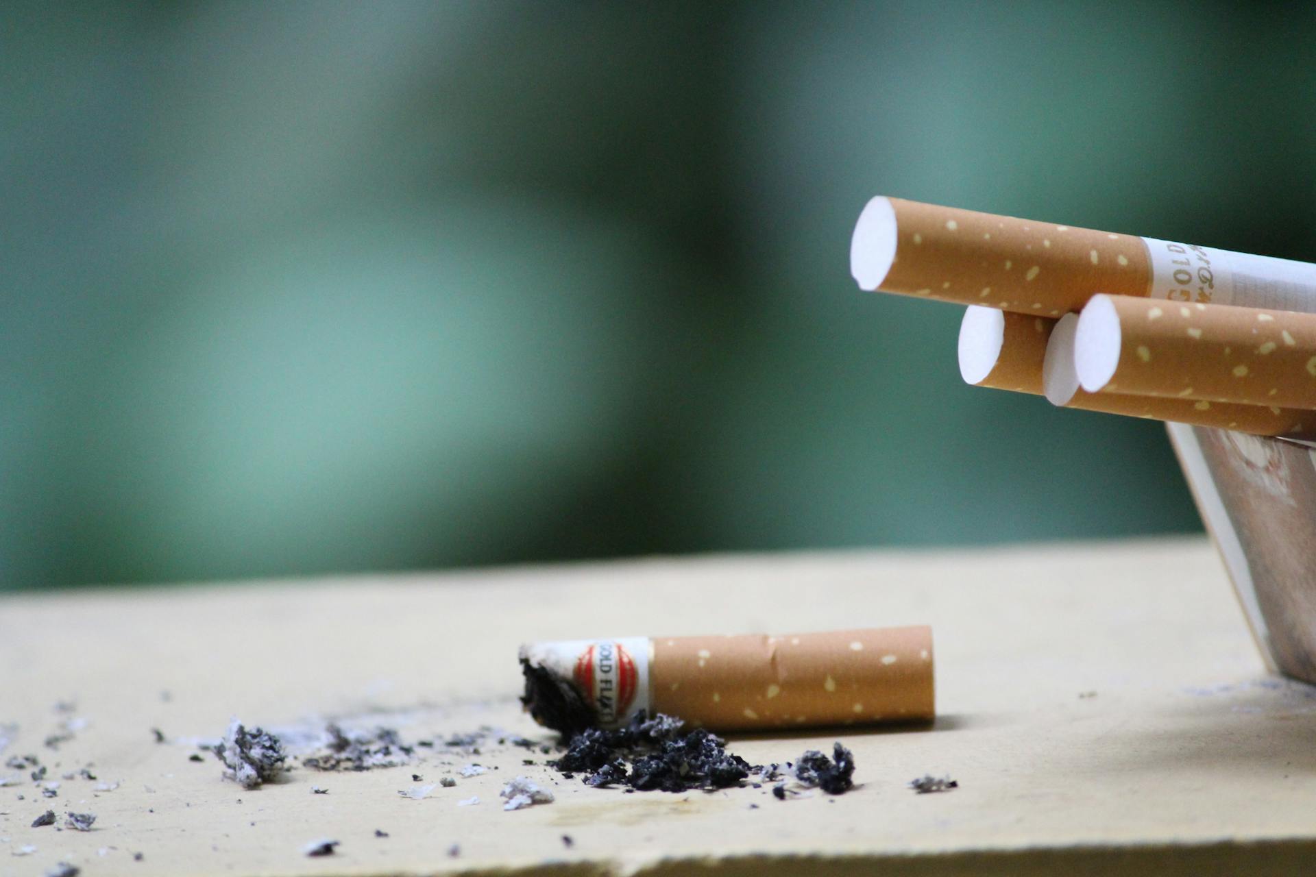
Tens of thousands of people are diagnosed in the States alone every year. The rates of incidence are even greater in countries where chewing tobacco and similar products are widespread, such as India, China, and Southeast Asia countries such as Myanmar, Indonesia, and Thailand.
The gold standard for diagnosing oral cancer, a biopsy, is stressful, invasive and painful, and costly too! The procedure, which includes pathology review, costs thousands of dollars to perform and results can take days to weeks to obtain. Someone must pay the bill, whether it be the patient and/or insurance provider. While, thankfully, greater than 90% of biopsies of suspicious oral lesions turn out to be negative, the anxiety-ridden waiting period and the cost involved could be reduced significantly if the clinician could be empowered to know a priori who truly needs a biopsy. Aaron Weinberg wants to empower the clinician to do just that.
At Case Western Reserve University, Aaron is developing a new way to test for oral cancer: the beta-defensin index (BDI). The lateral flow assays (LFAs) used to assess SARS-CoV-2 infections during the COVID-19 pandemic inspired his team to develop a way to determine whether someone has oral cancer using a non-invasive brush to collect cells. That led him to measure the concentrations of two beta defensin proteins in the lesion and unaffected tissue, both in the same patient. By calculating their ratios, he could identify patients with cancerous lesions at almost 100% sensitivity.
With such a test, people with lesions now have an affordable tool that clarifies if they should obtain a biopsy of their oral lesion.
Check out our interview with Aaron Weinberg below and read how he proposes to use his discovery to screen for oral cancer non-invasively while maintaining reproducibility.
PN: To start, what is oral cancer, and what got you so interested in studying cancers of the oral cavity?
AW: Oral cancer is a type of cancer that develops in the oral mucosa. Think of your tongue, your hard and soft palate at the roof of your mouth, and the inside lining of your cheeks and lips. All of that, and everything else that lines your oral cavity, comprises your oral mucosa. All of it works together to protect the underlying tissue from mechanical damage and invading microorganisms whenever you eat food, drink something, or speak. Oral cancer results from aberrant cells in the deepest layer of the mucosa that start proliferating uncontrollably, with the outcome resulting in a tumor.
You may have also noticed that whenever you bite your inner cheek, the bleeding stops shortly after the trauma. Your wound would heal quickly, and you can go on without worrying whether any bacteria will enter your bloodstream. It’s that amazing dynamic that drew me into studying proteins involved in innate protection of the oral cavity and how we can use them to screen for oral cancer.
PN: How is oral cancer currently diagnosed?
AW: Biopsy followed by a pathologist’s review is the gold standard for diagnosing oral cancers. Clinicians can perform either an excisional or an incisional biopsy at the affected site. Excisional biopsies remove the entire lesion, while incisional biopsies take a small portion of the lesional tissue. In a pathology lab, we will use microscopes to search for visual features that inform if the patient has a cancerous lesion. For example, we could look for abnormal cells in the mouth, known as epithelial dysplasia. We can also identify cells with abnormally large nuclei or nuclear polymorphisms; i.e., aberrant nuclear shapes, both characteristics of cellular atypia.
PN: Are there ways we could improve how we use biopsies to detect oral cancer?
AW: There is a lot of room for improvement when diagnosing oral cancers with biopsies. First, we must consider interrater variability in interpreting oral cancer. While different pathologists may interpret what a cancerous lesion looks like differently, they have made significant strides towards standardizing oral cancer diagnosis. Nevertheless, differences in interpretation remain, sometimes leading to disagreements in what constitutes a cancerous lesion.
Additionally, patients endure waits of several days to weeks while pathologists examine the biopsies underneath a microscope. That wait comes with a lot of stress and anxiety. Additionally, biopsies can lead to varying degrees of morbidity, such as pain, swelling and difficulty in eating, swallowing, and speaking if nerve damage occurs. Biopsies can, in some cases, also increase the risk of infection. When you perform a biopsy, you effectively create an open wound where oral bacteria can easily inhabit, grow, and enter the bloodstream.
Cost is another issue for patients who need a biopsy. In the States, a single biopsy followed by pathology review costs anywhere between $1200 to $3600, depending on the medical center. That’s a substantial cost, no matter who ends up paying for it.
PN: Give me some context here. What percentage of lesions tested turn out negative?
AW: In the US, 90% of query lesions that undergo biopsies end up being negative.
PN: That’s a lot of false alarms and thousands down the drain. But you’re not saying we should remove biopsies altogether, are you?
AW: I’m not saying we shouldn’t do biopsies anymore. Rather, I’m saying we should find ways to minimize the “false alarms.” Doing so would relieve stress and save money for people with lesions.
PN: That’s why you developed a supplementary approach based on beta defensins to detect lesions likely to be oral cancers. What are beta defensins, particularly the ones you used to assess oral cancer lesions?
AW: Defensins are a class of molecules known as antimicrobial peptides (AMPs). These are small proteins that can kill microbes, whether they be bacteria, fungi, or encapsulated viruses.
Defensins can be classified as alpha or beta defensins. While cells across the body express alpha defensins, only epithelial cells produce beta defensins. Our oral mucosa is full of these epithelial cells. There, beta defensins can kill microbes directly, but they can also attract immune cells from the bloodstream to any area where your oral cavity is susceptible to infection. It’s this combination of recruiting immune cells and killing microbes directly that prevents an oral wound from being infected. That includes biting your tongue and your cheek.
PN: We’ve identified situations where beta defensins would be upregulated, but how do our cells regulate beta defensin expression?
AW: Our cells express special proteins called transcription factors. They tell the genes in our cells to start producing certain proteins. We discovered that two of the human beta defensins (HBD), HBD-2 and HBD-3, are regulated by two distinct transcription factors. HBD-2 is regulated by NF-kB, a transcription factor essential for activating several regulatory cascades related to inflammation. On the other hand, HBD-3 is regulated by epidermal growth factor receptor (EGFR). This receptor triggers a series of biological processes that promote epithelial cell proliferation and movement to help in wound healing.
PN: So, two distinct regulatory processes control HBD-2 and HBD-3 expression. I believe it’s these two molecules that inspired the HBD index, or BDI, for detecting oral cancer.
AW: That’s right. Since HBD-3 expression is linked to cell proliferation, I surmised that uncontrolled proliferation would be induced by overexpressing EGFR, leading to a lot of HBD-3 in early-stage oral cancer lesions. Through several techniques, we showed that oral squamous cell carcinomas had high HBD-3 levels compared to HBD-2 in the cancerous lesion. We then surmised that the ratio of HBD-3/HBD-2 proteins in cells obtained by brushing suspicious oral lesions, compared to the contralateral normal site in the same patient, could distinguish oral cancer from non-cancerous lesions. We refer to this ratio as the Beta-Defensin Index (BDI). After conducting a proof of principle study, followed by blinded discovery and validation studies, we found that we could indeed distinguish oral cancer from noncancerous lesions accurately and reproducibly.
PN: How would you measure BDI towards screening for oral cancers?
AW: We are currently taking samples to the lab, where the enzyme-linked immunosorbent assay, or ELISA is performed. This assay uses antibodies that bind specifically to HBD-2 and HBD-3. By the end of the assay, we would get readings based on how much of the antibodies bind to HBD-2 and HBD-3. If there is more antibody binding to the molecule, we would know that more of the specific beta defensin will be present.
Amid our experiments, we found that people we sampled whose oral mucosa was healthy had HBD-3/HBD-2 ratios of around 1:1. However, patients who had oral cancer lesions tended to have BDIs scores of at least 1.25:1 up to 30:1. What’s amazing is that this metric provided us with nearly 100% sensitivity for identifying oral cancer lesions in our patient population. As for specificity, we found a false positive rate of just 17%. The false positives arose from us picking up precancerous lesions. This ends up being beneficial for a clinician, since it gives a chance to warn patients early about a precancerous lesion.
PN: You’ve done your experiments in the lab, but could we perform the test faster, perhaps similar to what we did with COVID-19 test kits?
AW: Absolutely. We’re currently developing a lateral flow assay (LFA) that measures the abundances of both beta defensins in minutes. The rapid assay would comprise two components where HBD-2 and HBD-3 would be quantified from the normal oral mucosa and the lesion. This would give us a much cheaper approach for quantifying these proteins and remove waiting times that arise from shipping samples to the lab for further testing.
Right now, we’re developing a kit with our collaborators at Hemex Health. They have experience quantifying proteins with LFAs, having done so for sickle cell disease (SCD) and beta thalassemia. Our intention will be to go through the process of receiving FDA approval for our LFA. Should that happen, we will then be able to begin commercializing the test in the States and eventually the rest of the world.
PN: How are you hoping to make the BDI more widely adopted for screening oral cancer?
AW: First, we’re not replacing biopsies with the BDI. Our intent is to use the BDI as a screening tool by letting people know if their oral lesions are likely to be cancerous and if they should have a biopsy done. Currently, we are building on several collaborations with colleagues in India, Taiwan, and the ASEAN nations where oral cancer has grown to epidemic proportions. We recently attended a PORTENT-held conference in Bangalore. There, we presented our groundbreaking work and opened the doors for future integration into the Gazelle platform developed by Hemex Health.
It became clear to me while working with our various collaborators, including those at Queen Mary’s University Hospital in East London, that refugees from these nations are at higher risk of oral cancer than populations in Western countries. This is primarily due to the habitual consumption of tobacco and betel nut; customs they brought with them from the subcontinent and SE Asia. These habits drive lesion formation owing to the carcinogens found in these products.
The billions of people who live across Asia give us a huge opportunity to alleviate the hardships that come with oral cancer. With ongoing collaborations, I’m hopeful that we can solidify and standardize the non-invasive BDI rapid test as a viable metric for detecting oral cancer. I don’t want the test to be available just in the West. I also want it to reach people living in low- and middle-income countries, where laboratory/pathology services are oftentimes lacking.