Share this article


Help your customers see the core of your life's work.
Let us translate your science and bring your marketing to life!
The life sciences industry has developed many approaches to preparing new disease therapeutics. More and more, modern biomedical research seeks to alter how cells behave by introducing functional molecules into them. These can range from nucleic acids such as mRNA or plasmid DNA to larger protein complexes such as CRISPR-Cas9 ribonucleoproteins. With so many new kinds of payloads being developed, many research groups have developed new ways to deliver them into target cells, such as transfections.
Historically, scientists have used various physical approaches to perform transfections such as electroporation. With electroporation, they would briefly shock the cells to form transient pores that are big enough and appear just long enough to introduce the cargo into the cells. The problems with inducing cell stress, however, have encouraged researchers to develop new approaches to transfect cells. In fact, I talked about one such (include button to interview). Yet with new transfection approaches, one question continues to persist:
How do you know that the transfection successfully delivered the cargo?
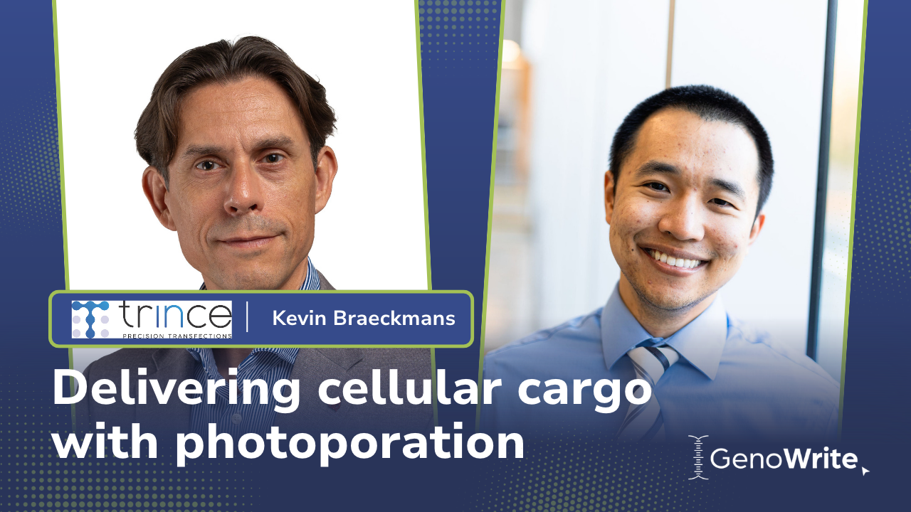
I had a great opportunity to speak with Kevin Braeckmans, CEO and co-founder of Trince, about this very question. His team developed the LumiPore® platform, which uses a laser-based instrument and nanosensitizers to gently open transient pores in the cell membranes of recipient cells. These short-lived openings let external molecules enter the cell with high efficiency, while preserving the cell’s health and function. It was through the development of this approach that Trince won the prestigious nature Spinoff Prize in 2024 and the SLAS Ignite Award at SLAS Europe 2026.
As you’ll see from our interview, addressing this very question is a core aspect of securing adoption of new transfection technologies like photoporation into therapeutic R&D. Not only that, but his platform can also perform transfections directly on adherent cells at scale. Check it out below!
PN: Your new website mentions that scientists can now conduct transfections without needing to detach the cells from their in situ configurations, and at high-throughput rates no less. How did you make this possible?
KB: The LumiPore® workflow itself is simple. LumiSense® nanosensitizers are added directly to cells growing in standard culture vessels and together with the payload of interest. The sample is then placed in the LumiPore® device, where they will receive a brief laser treatment. Within minutes, the payload will have been delivered and the cells will remain in excellent condition, ready for downstream analysis or further processing.
Because cells do not need to be transferred to dedicated transfection vessels or media, the process can be readily applied to both suspension and adherent cells. Scientists can perform transfections directly in their preferred culture vessels; the only requirement is a light-transparent bottom, which is standard for the vast majority of cell cultureware. To support high-throughput drug-discovery workflows, the technology is compatible with high-density culture plates, including 384- and even 1,536-well formats.
PN: What kinds of cells and experiments would most benefit from being able to transfect cells while keeping them adherent in their existing culture format?
KB: Detaching adherent cells, whether by trypsinisation or by other enzymatic or mechanical methods, is a significant stressful event in itself: it cleaves surface proteins and breaks the contacts that cells have formed with each other and with their culture surface. For sensitive adherent cell types such as macrophages, stem cells, or iPSC-derived neurons and cardiomyocytes, lifting them off their culture surface destroys the morphology and connections they have built up, such as neurite networks in neurons or synchronised beating in cardiomyocyte monolayers. After transfection in suspension, the cells then have to be reseeded, optionally after sorting to enrich for successfully transfected cells, and given time to reattach and recover. Not all of them will, and for some cell types, such as mature neurons, re-plating is hardly feasible at all.
Each of these steps causes delays, adds stress and can alter the cells’ morphology, adhesion and gene expression. Yet it is precisely these features, their shape, their contacts and the functions that depend on them, that make these cells meaningful to study in the first place. If the workflow alters them, you are no longer modelling the cell behaviour you set out to study.
These cell types are important targets of cell and gene therapy, regenerative medicines, and genetic therapies. A transfection protocol that keeps the cells intact with minimal changes to their phenotypes would help researchers evaluate the efficacy of these therapies in conditions similar to those observed in vivo. If you can transfect them exactly where they’re growing, without ever moving them, you avoid the pain of altered phenotypes that can compromise your data.
PN: An ideal transfection efficiency is usually cited as 70-100%. If transfection efficiencies fall below that, what can cause it?
KB: We have to first begin by defining what makes a successful transfection. Any successful transfection requires two changes in the cell to be observed:
Have the cells taken up the cargo?
Do the cells express the expected change in phenotype with the cargo?
This distinction is especially important to note when employing any transfection protocol, including photoporation. Low transfection efficiency can be caused either by inefficient cargo delivery or by the cells’ inability to process the delivered cargo effectively. With photoporation, inefficient cargo delivery is rarely the cause, whereas the cell’s ability to process the cargo depends on its biology.
PN: In light of what you’ve just said, what example can you give of cell biology resisting transfections?
KB: Recent evidence from our research has shown that photoporation delivers macromolecules into naive T cells just as efficiently as into activated T cells. However, what we found was that activated T cells are consistently more likely to express the phenotype of interest, whether it’s protein expression or a gene knockout. This is most likely because naive T cells sit in a quiescent state, with low metabolic and transcriptional activity. How active the cells are at the time of transfection, then, affects the final outcome.
PN: Sounds like this logic can also apply to the nature of the payload itself.
KB: Yes, that’s right. The design and quality of the payload equally determines the eventual outcome. We have had customer cases where a plasmid design was suboptimal, leading to markedly lower expression than reference plasmids tested on the same cell type. We have also seen CRISPR knockout experiments where the targeted gene in the chosen cell type happened to be less accessible, resulting in lower efficiency than expected based on reference measurements targeting more accessible genes. In both cases, the delivery step worked fine. The shortfall came from something downstream of it.
PN: It also stands to reason that these factors are common not just to photoporation, but to other delivery methods, too, no?
KB: Correct. Aside from the payload quality and the cell type, we also have to consider how the construct was designed and how accessible a target gene or locus is in a given cell type. Each will affect transfection outcomes regardless of how the payload entered the cell.
PN: We’ve spent time discussing the factors that affect transfection efficiency outside of photoporation. What factors could we control with the photoporation protocol itself?
KB: There are two parameters specific to the photoporation itself that affect how many cells survive and successfully took up the cargo after the transfection:
We provide a straightforward, generic but flexible protocol for tuning both, so users can quickly establish the right settings whenever they switch between cell types or applications. In practice, this optimization is usually done with a fluorescently labeled model molecule of similar size to the actual payload of interest. That lets us confirm successful delivery independent of the expected biological outcome and gives a clear starting point for troubleshooting if that outcome does not show up as expected.
PN: How have these insights affected how you describe your technology?
KB: You may have noticed that we describe our technology as an intracellular delivery platform rather than simply a transfection technology. Our data demonstrates that delivery efficiency is not always coupled to the outcome of the transfection. Treating them as one and the same makes troubleshooting a transfection protocol that much harder.
PN: Ensuring that cells behave as they should after treatment is a major talking point when it comes to transfections. How should a scientist confirm whether the cells still behave as expected after treatment?
KB: Using an intracellular delivery technology that preserves a cell’s phenotype and functionality is extremely important for getting reliable results from that comparison. That’s why we need controls to ensure that the cells still behave as expected after treatment.
The general approach is to compare treated cells against an untreated control on the specific readout that matters for your application. The details vary depending on the cell type you are working with: checking for changes in gene expression at the transcriptome or protein level, measuring growth rate, or running a functionality assay specific to that cell type. That could mean measuring how efficiently cytotoxic T cells kill cancer cells and assessing whether iPSCs can differentiate or have differentiated into the desired somatic cell type, or looking at the migration speed of mesenchymal stromal cells, just to give a few examples.
Regardless of the approach, any research team will need to know intimately their cell’s behaviours and what phenotypes to monitor before and after a transfection.
PN: Looking ahead, what kinds of preclinical drug development efforts do you think would benefit most from adopting photoporation?
KB: There are four major areas in early-stage drug discovery where we see real potential for photoporation to be adopted:
Altogether, we see three benefits from implementing photoporation for scientists in drug discovery.
That combination of a simpler workflow, more biologically relevant outcomes, and scalability across automated high throughput formats is the core value we aim to bring to preclinical drug development. Ultimately, we hope this enables earlier, more predictive data on how drug candidates will behave in humans, addressing one of the persistent challenges in drug development.

Paul Naphtali is a seasoned online marketing consultant. He brings to the table three years of online marketing and copywriting experience within the life sciences industry. His MSc and PhD experience also provides him with the acumen to understand complex literature and translate it to any audience. This way, he can fulfill his passion for sharing the beauty of biomedical research and inspiring action from his readers.
Help your customers see the core of your life's work.
Let us translate your science and bring your marketing to life!